
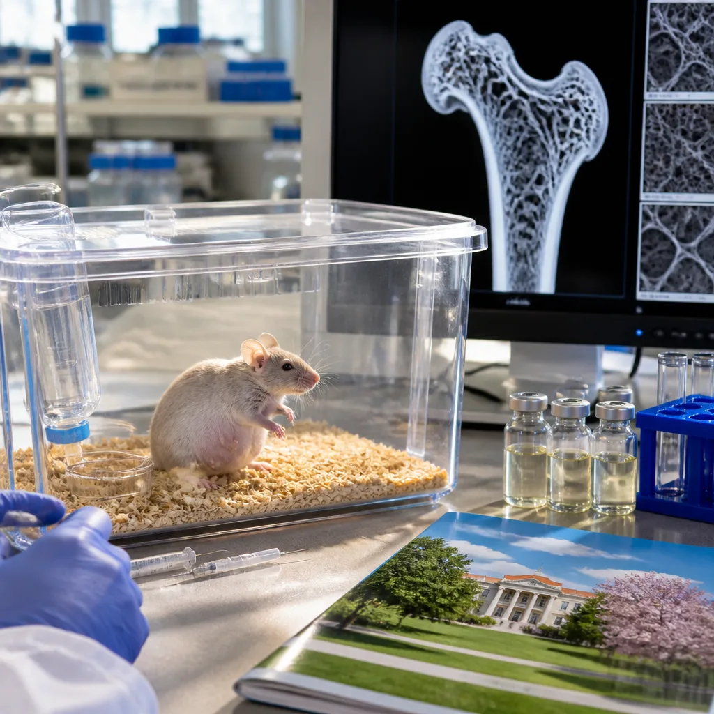
Researchers at the University of Missouri report that reducing activity of the protein myostatin strengthened bone structure and increased muscle mass in a preclinical mouse model of osteogenesis imperfecta (OI) during pregnancy, a period when women typically lose bone density and when many standard OI drugs are not recommended.
Key findings from the study
- Lowering myostatin improved bone density and altered bone architecture in pregnant mice with OI, helping bones better resist stress without changing the underlying collagen defect.
- The intervention increased muscle mass in the animal model and did not appear to harm the mother or offspring in the experiments reported.
- Investigators propose that modifying muscle-related biology can change bone structure enough to reduce fracture risk even when the bone material itself remains genetically determined.
The study addresses a particular clinical challenge: pregnancy and breastfeeding are periods of accelerated maternal bone loss because fetal and neonatal calcium demands increase. That physiologic loss can be especially hazardous for women with OI, a genetic connective tissue disorder that weakens bone due to defective collagen. In addition, many conventional OI therapies are avoided in pregnancy, limiting treatment options.
"Pregnancy and breastfeeding are already natural periods of bone density loss for women, as a baby's growth has a large calcium demand both inside and outside the womb,"
Study authors described OI bone as having inherently weaker material properties compared with healthy bone. They used an analogy to emphasize their approach: rather than changing the bone’s material, they increased the amount and altered the structure to make it harder to break. As the senior author put it, boosting structural quantity and geometry can compensate, at least partially, for inferior material quality.
| Concept | Implication in study |
|---|---|
| Bone material quality | Unchanged (genetically determined in OI) |
| Bone structure/quantity | Improved by lowering myostatin activity in mice |
Context and potential consequences
The findings point to a therapeutic angle that targets muscle–bone interactions: myostatin normally limits muscle growth, and its reduction can enlarge muscle mass and influence the forces applied to bone, prompting structural adaptation. If these mechanisms translate to humans, modulation of myostatin or related pathways could offer a strategy to protect pregnant women with OI and other conditions that make post-partum bone recovery difficult.
Important caveats remain. The work reported is preclinical, conducted in a mouse model. Translating animal results into safe, effective human therapies requires extensive additional study, including evaluation of long-term outcomes and reproductive safety. The authors note that, in their experiments, lowering myostatin did not appear to harm mothers or offspring — an encouraging but preliminary observation.
Osteogenesis imperfecta encompasses more than 10 genetic types and ranges in severity. Because the disorder affects the fundamental composition of bone, most treatments aim to reduce fracture risk through supportive measures, physical therapy and — when appropriate — pharmacologic agents. During pregnancy, treatment choices are constrained, so new approaches that are safe in that window would fill an important clinical gap.
Future research priorities include repeating findings in additional models, mapping molecular pathways that link muscle changes to bone architecture, and designing carefully controlled clinical trials to assess safety and efficacy in humans. For now, the study adds to accumulating evidence that targeting muscle-regulatory proteins can have meaningful effects on skeletal health.
Implications: The research suggests a promising direction for protecting skeletal integrity in pregnant individuals with OI and possibly other women at risk of prolonged bone loss, but human studies are needed before clinical application.